


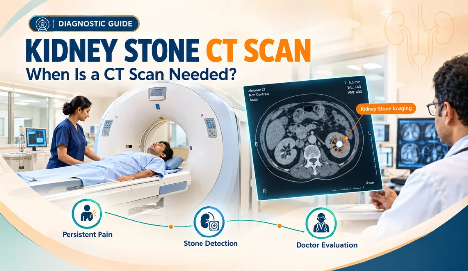
Kidney Stone CT Scan: When Is a CT Scan Needed?
Kidney stones can cause sudden and severe pain, blood in the urine, nausea, vomiting and difficulty passing urine. When these symptoms occur, doctors may recommend imaging to determine whether a stone is present, where it is located and whether it is blocking the urinary tract. Among the available imaging tests, a CT scan can provide detailed information about the size, location and number of kidney stones.
However, not every person with suspected kidney stones automatically needs a CT scan. The choice of imaging depends on symptoms, age, pregnancy status, previous medical history, suspected complications and the information already available from other tests such as ultrasound.
Understanding when a CT scan is useful can help patients avoid unnecessary investigations while ensuring that important complications are detected at the right time.
Table of Contents
1. What Is a Kidney Stone CT Scan?
2. Why Is a CT Scan Used for Kidney Stones?
3. When Is a CT Scan Needed for Kidney Stones?
4. CT Scan vs Ultrasound for Kidney Stones
5. What Can a CT Scan Show?
6. Is CT Scan Necessary for Every Kidney Stone Patient?
7. When Should You See a Specialist?
8. What Happens After a Kidney Stone CT Scan?
What Is a Kidney Stone CT Scan?
A CT scan, or computed tomography scan, uses X-rays and computer processing to create detailed images of the urinary system. When kidney stones are suspected, doctors may use a specialised CT examination of the kidneys, ureters and bladder, commonly referred to as a CT KUB.
A non-contrast CT scan is frequently used for suspected urinary stones because many stones can be clearly identified without the need for contrast dye. It can provide detailed information about the stone and surrounding urinary structures.
Unlike a basic X-ray, CT imaging can detect many stones that are difficult to see on a standard abdominal X-ray and can provide much more precise information about their location and size.
Why Is a CT Scan Used for Kidney Stones?
The main purpose of CT imaging is to answer several important questions:
• Is there actually a kidney or ureteral stone?
• How large is the stone?
• Where exactly is the stone located?
• Is the stone blocking urine flow?
• Is there swelling of the kidney or urinary tract?
• Are there multiple stones?
• Is there another condition causing the patient's pain?
This last point is particularly important. Severe abdominal or flank pain is not always caused by a kidney stone. Other urinary, gastrointestinal, gynaecological or abdominal conditions can produce similar symptoms. Detailed imaging can help doctors investigate alternative causes when the diagnosis is uncertain.
When Is a CT Scan Needed for Kidney Stones?
A CT scan may be recommended when the diagnosis is uncertain, when symptoms are severe or unusual, or when the doctor needs detailed information about the stone before deciding on treatment.
Common situations where CT imaging may be considered include:
| Situation | Why CT May Be Helpful |
|---|---|
| Severe flank or abdominal pain | Can help identify a stone and determine whether it is causing obstruction. |
| Uncertain diagnosis | Helps distinguish kidney stones from other causes of abdominal or urinary pain. |
| Suspected ureteral obstruction | Can show the stone's exact location and associated urinary tract changes. |
| Persistent symptoms | May provide more detailed information when symptoms continue despite initial assessment. |
| Before certain stone treatments | Detailed stone size and location can help the urologist plan appropriate treatment. |
| Multiple or recurrent stones | Can provide a detailed assessment of the overall stone burden. |
Is CT Scan Needed for Every Kidney Stone Patient?
No. The decision depends on the patient's individual circumstances.
For some patients, especially those with a known history of kidney stones and typical symptoms, an ultrasound or another appropriate imaging test may provide sufficient initial information. In other cases, CT may be recommended because the doctor needs greater detail or wants to rule out another potentially serious condition.
The goal is to select the imaging test that provides the necessary diagnostic information while taking factors such as radiation exposure into consideration.
CT Scan vs Ultrasound for Kidney Stones
Ultrasound and CT scans are both useful in the evaluation of kidney stones, but they provide different types of information.
| Feature | Ultrasound | CT Scan |
|---|---|---|
| Radiation | Does not use ionising radiation | Uses X-rays |
| Stone detection | Can detect many kidney stones and urinary obstruction, but small or ureteral stones may be difficult to visualise | Highly detailed assessment of urinary stones and their location |
| Stone size | Can provide an estimate, but measurements may be less precise in some situations | Provides detailed measurement and stone burden assessment |
| Kidney swelling | Can identify hydronephrosis or urinary tract dilation | Can provide detailed information about obstruction and surrounding structures |
| Pregnancy | Often preferred as an initial imaging method | Generally avoided unless the clinical situation requires careful consideration of CT imaging |
Ultrasound is particularly valuable because it does not expose the patient to radiation. However, CT can provide more detailed information when a precise diagnosis is required.
Why Is CT Often More Detailed Than Ultrasound?
Ultrasound works using sound waves and can provide useful information about the kidneys, bladder and urinary tract. However, the ability to visualise a stone can be affected by the stone's location, patient anatomy and bowel gas.
CT imaging can produce cross-sectional images of the urinary tract and is generally better at identifying the exact location and size of many urinary stones. It can also provide information about the surrounding abdominal structures.
This detailed information can be particularly valuable when the symptoms are severe but the diagnosis is uncertain or when treatment planning requires an accurate assessment of the stone.
What Is a Non-Contrast CT KUB?
A non-contrast CT KUB is a CT scan focused on the kidneys, ureters and urinary bladder that is performed without intravenous contrast dye. It is commonly used for evaluating suspected urinary stones because many stones are clearly visible without contrast.
The scan can help identify the size and location of a stone and whether there are signs of urinary obstruction.
Whether a non-contrast CT KUB is appropriate depends on the patient's symptoms and clinical situation. The treating doctor or radiologist determines the most suitable imaging protocol.
Can a CT Scan Show the Exact Size of a Kidney Stone?
CT imaging can provide a detailed measurement of kidney and ureteral stones. Knowing the approximate size is important because stone size can influence the likelihood of natural passage and the choice between observation and active treatment.
For example, a small stone may be managed differently from a large stone. However, stone size should never be considered in isolation. Location, obstruction, infection, kidney function and symptoms are also important.
Can CT Show Whether a Kidney Stone Is Blocking Urine?
Yes. CT imaging can provide important information about urinary obstruction and changes in the urinary tract associated with a stone.
A stone that blocks the ureter can prevent normal urine drainage from the kidney to the bladder. This may result in swelling of the urinary collecting system and can contribute to significant pain.
Obstruction becomes particularly concerning when it occurs together with infection or impaired kidney function. Such situations require prompt medical assessment.
When Is CT More Important Before Kidney Stone Treatment?
Detailed imaging can be particularly useful when a procedure is being considered. The urologist may need to know the stone's exact location, size, number and characteristics before deciding between observation, ESWL, ureteroscopy or PCNL.
For larger stones, multiple stones or complex stone burdens, imaging plays an important role in treatment planning. The information obtained from the scan can help the specialist choose an approach that offers an appropriate balance between effectiveness and safety.
Can CT Detect Other Problems Besides Kidney Stones?
One of the advantages of CT imaging is that it can provide information about structures beyond the urinary tract. This can be useful when symptoms are not clearly explained by a kidney stone.
Severe abdominal or flank pain can have many possible causes. Depending on the imaging protocol and clinical situation, CT may help doctors investigate other abdominal or urinary conditions when appropriate.
However, CT should not be treated as a substitute for clinical examination. The scan findings need to be interpreted together with symptoms, physical examination, urine tests, blood tests and medical history.
Is a CT Scan Safe for Kidney Stone Diagnosis?
CT scans use ionising radiation, so doctors generally aim to use them when the expected diagnostic benefit justifies the exposure. Modern CT technology and appropriate protocols can help reduce radiation exposure while maintaining useful image quality.
The decision is particularly important for younger patients, people who may require repeated imaging and pregnant patients. In such situations, the doctor may consider ultrasound or other approaches when clinically appropriate.
What About CT Scans During Pregnancy?
Pregnancy requires special consideration because CT uses ionising radiation. Ultrasound is generally preferred as the initial imaging method when kidney stones are suspected during pregnancy.
If symptoms are severe, the diagnosis remains uncertain or complications are suspected, the medical team may consider additional imaging after carefully weighing the benefits and risks. Pregnant patients should always inform the doctor and radiology team about their pregnancy or possible pregnancy before imaging.
What Tests May Be Done Along With a Kidney Stone CT Scan?
CT imaging is only one part of kidney stone evaluation. Doctors may also recommend urine and blood investigations to understand the patient's condition and identify possible complications.
| Test | What It May Help Assess |
|---|---|
| Urine examination | Blood in urine, signs of infection and other urinary abnormalities |
| Urine culture | Whether bacteria are present when urinary infection is suspected |
| Blood tests | Kidney function and other relevant health parameters |
| Ultrasound | Kidney stones, bladder findings and urinary tract swelling |
| CT KUB | Detailed assessment of stone size, location, number and obstruction |
When Should You Consult a Kidney Stone Specialist?
If you have severe or recurring flank pain, blood in the urine, repeated urinary symptoms or a previous history of kidney stones, medical evaluation can help determine whether imaging is required.
A specialist may recommend CT imaging when the diagnosis needs clarification or when detailed information is required for treatment planning. Consult a Specialist for Accurate Kidney Stone Diagnosis rather than relying only on symptoms or assuming that every episode of abdominal pain is caused by a kidney stone.
What Happens After the CT Scan?
After the scan, the radiologist interprets the images and prepares a report. The treating doctor or urologist then combines the imaging findings with the patient's symptoms, examination and laboratory results.
If a stone is identified, the next decision may be observation, medical management, follow-up imaging or active treatment. The appropriate option depends on the stone's size and location as well as whether there is obstruction, infection or another complication.
How to Read a Kidney Stone CT Scan Report
Receiving a CT scan report after experiencing kidney stone symptoms can be confusing. Medical terms such as calculus, ureteric calculus, hydronephrosis, obstruction, renal calculus and HU may appear in the report without clearly explaining what they mean for the patient.
A CT report provides important information, but the report should always be interpreted together with your symptoms, urine and blood tests, medical history and clinical examination. A particular finding does not automatically mean that surgery is required.
What Information Does a Kidney Stone CT Report Usually Include?
A detailed CT report may describe the number of stones, their size, exact location, density, whether urine flow is obstructed and whether there is swelling of the kidney or ureter.
| CT Report Finding | What It Generally Means |
|---|---|
| Renal calculus | A stone is present within the kidney. |
| Ureteric calculus | A stone is present within the ureter, the tube carrying urine from the kidney to the bladder. |
| Stone size | Provides an estimate of the stone's dimensions and helps assess treatment options. |
| Hydronephrosis | Swelling or dilation of the kidney's urine-collecting system, often associated with impaired urine drainage. |
| Hydroureter | Dilation of the ureter, which may occur when urine flow is obstructed. |
| Obstruction | Indicates that urine drainage may be impaired. |
| HU / Hounsfield Unit | A measurement of CT density that can provide information about the stone and may help with treatment planning. |
What Does “Renal Calculus” Mean in a CT Report?
The word calculus simply means a stone. Therefore, a “renal calculus” means a kidney stone located inside the kidney.
A report may mention one or more calculi and provide their approximate measurements in millimetres. It may also specify which kidney is involved and which part of the kidney contains the stone.
A kidney stone that remains inside the kidney may behave differently from a stone that has moved into the ureter. This distinction is important because ureteral stones are more likely to cause acute obstruction and renal colic.
What Does “Ureteric Calculus” Mean?
A ureteric calculus is a stone located in the ureter. Because the ureter is relatively narrow, a stone can become lodged and interfere with normal urine drainage.
This can cause sudden, severe pain that may begin in the side or back and radiate toward the lower abdomen or groin. Some patients may also experience nausea, vomiting or blood in the urine.
The exact location of a ureteral stone is important because the likelihood of spontaneous passage and the choice of treatment can change as the stone moves through the urinary tract.
What Does Hydronephrosis Mean on a CT Scan?
Hydronephrosis means dilation or swelling of the kidney's collecting system. When a stone blocks urine flow through the ureter, urine can build up behind the obstruction and cause this dilation.
The presence and severity of hydronephrosis can provide important information to the treating doctor. Mild dilation does not necessarily mean emergency surgery, but significant or worsening obstruction requires appropriate medical evaluation.
Hydronephrosis associated with infection, severe symptoms or impaired kidney function is more concerning and may require urgent treatment.
What Does Hydroureter Mean?
Hydroureter refers to dilation of the ureter. It may occur when urine cannot flow normally toward the bladder because of an obstruction.
When a CT report mentions both a ureteric stone and hydroureter, the findings may suggest that the stone is affecting urine drainage. The significance depends on the severity of obstruction and the patient's symptoms and laboratory results.
Why Is Stone Location Important on a CT Scan?
Knowing the exact location of a stone can help the urologist determine whether observation is reasonable or whether active treatment should be considered.
| Location | Possible Significance |
|---|---|
| Kidney | May remain asymptomatic or may cause recurrent pain, infection, bleeding or future obstruction. |
| Upper ureter | Can obstruct urine drainage and cause significant flank pain. |
| Middle ureter | Passage depends on size, anatomy and stone progression. |
| Lower ureter | Some stones may have a greater opportunity to reach the bladder, but size and symptoms remain important. |
| Ureterovesical junction | The area where the ureter meets the bladder; stones may become lodged here. |
Does CT Stone Size Determine Whether Surgery Is Needed?
Stone size is one of the most important factors, but it does not independently determine whether surgery is necessary.
In general, very small stones have a greater possibility of passing naturally. As stone size increases, spontaneous passage becomes less predictable. Larger stones are more likely to require active treatment.
However, a smaller stone can still require urgent treatment if it causes infection with obstruction, severe uncontrolled symptoms or significant impairment of kidney function.
What Does Stone Density Mean on a CT Scan?
CT scans can provide information about the density of a kidney stone, often expressed in Hounsfield Units (HU). Stone density can provide clues about how the stone may respond to certain treatment methods.
For example, some stones may be harder and less responsive to shock-wave fragmentation. The CT appearance and density can therefore be one of several factors considered when a urologist evaluates whether ESWL, ureteroscopy or another treatment would be appropriate.
However, HU should not be interpreted as a simple “hard stone” or “soft stone” label. Stone composition, CT technique and other factors can affect the measurement.
Can CT Tell What Type of Kidney Stone You Have?
CT findings can sometimes provide clues about the likely characteristics of a stone, but CT alone cannot always determine its exact chemical composition.
If a stone is removed or passed, laboratory stone analysis can provide more direct information about its composition. This can be particularly useful for patients who repeatedly develop kidney stones because prevention strategies can be tailored to the type of stone.
What Does “No Hydronephrosis” Mean?
If a CT report says there is “no hydronephrosis,” it generally means the scan did not show significant dilation of the kidney's collecting system at the time of imaging.
This can be reassuring, but it does not mean that a kidney stone is harmless or that treatment can never be required. A stone may still cause pain, grow over time, move into the ureter or cause other problems.
What Does “Mild Hydronephrosis” Mean?
Mild hydronephrosis means that there is a small degree of dilation of the urinary collecting system. It may occur when a stone partially interferes with urine drainage.
The finding needs to be interpreted in context. The location and size of the stone, symptoms, kidney function and presence or absence of infection all influence the next step.
What Does “Obstructing Calculus” Mean?
An obstructing calculus is a stone that is interfering with the normal flow of urine. The obstruction may be partial or significant depending on the situation.
An obstructing stone can cause pain and swelling of the urinary system. If obstruction is accompanied by infection, it can become a serious medical problem requiring urgent attention.
Kidney Stone CT Findings That May Require Prompt Medical Attention
Certain combinations of CT findings and symptoms should be taken seriously. These may include an obstructing stone with fever, significant urinary tract dilation with worsening kidney function, inability to pass urine or severe symptoms that cannot be controlled with prescribed treatment.
Patients should not wait for a routine appointment when severe pain is accompanied by fever or chills, confusion, marked weakness or other signs of serious infection.
Why CT Findings Must Be Combined With Blood and Urine Tests
A CT scan provides anatomical information, but it does not provide the complete picture of a patient's health.
Urine testing can help identify blood or signs of infection, while urine culture may be recommended when infection is suspected. Blood tests can help assess kidney function and other factors that may influence treatment decisions.
This combination of imaging and laboratory evaluation is particularly important when an obstructing stone is suspected.
Can a CT Scan Show Multiple Kidney Stones?
Yes. CT imaging can identify multiple stones in the kidneys and urinary tract and can help estimate the overall stone burden.
This is important because treatment may be different for one small stone compared with several stones or a large collection of stones. The urologist may need to consider whether all stones require treatment or whether some can be monitored.
Can a CT Scan Detect a Small Kidney Stone?
CT is highly sensitive for many urinary stones, including stones that may be difficult to identify on other imaging methods. However, the ability to detect a particular stone can depend on its size, composition and the imaging technique used.
A very small finding should also be interpreted carefully because not every tiny calcification near the urinary tract necessarily represents a clinically significant stone.
When Is Ultrasound Enough Instead of CT?
Ultrasound may be sufficient in selected patients, particularly when the clinical situation is straightforward and the doctor mainly needs to assess the kidneys, bladder or presence of urinary tract dilation.
Ultrasound is also particularly valuable when avoiding radiation is important, such as during pregnancy.
However, if symptoms are severe, the diagnosis is uncertain, a ureteral stone is suspected or detailed stone mapping is needed for treatment planning, the doctor may recommend CT imaging.
Should You Get a CT Scan Every Time You Have Kidney Stone Pain?
Not necessarily. Repeated CT scans should be based on clinical need rather than performed automatically every time symptoms occur.
A doctor may consider previous imaging, current symptoms, physical examination, urine and blood test results and whether there is a change in the patient's condition before deciding whether repeat imaging is necessary.
When follow-up imaging is needed, the most appropriate test depends on the clinical situation and the information the doctor needs to obtain.
What Should You Do After Receiving Your CT Report?
Do not try to determine your treatment only from the stone measurement written in the report. A 6 mm, 8 mm or 10 mm stone can have very different implications depending on whether it is inside the kidney or blocking the ureter.
Take the CT report and, when available, the actual imaging study to a qualified urologist. The specialist can correlate the imaging with your symptoms and laboratory findings and determine whether observation, medication, follow-up imaging or active stone treatment is appropriate.
When Should You Consult a Specialist?
You should consider specialist evaluation if your CT scan shows a ureteral stone, significant stone burden, obstruction, hydronephrosis, multiple stones or a stone that is not passing as expected.
Specialist assessment is also important when you have repeated kidney stones or when the report contains findings that you do not understand. Consult a Specialist for Accurate Kidney Stone Diagnosis so the CT findings can be interpreted in the context of your complete medical condition.
What Happens If the CT Scan Shows a Large Kidney Stone?
A large stone does not necessarily mean that emergency surgery is required, particularly when it is sitting inside the kidney without infection or obstruction. However, larger stones are less likely to pass naturally and may require planned active treatment.
The urologist may consider procedures such as ESWL, ureteroscopy or PCNL depending on the stone's size, location, density, anatomy and overall stone burden.
For very large or complex stones, PCNL may be an important treatment option because it allows the surgeon to directly access and remove a substantial amount of stone material.
Why You Should Not Ignore a Kidney Stone Found on CT
Some kidney stones can remain stable for a long time without causing symptoms, but others may grow, move into the ureter, cause obstruction or contribute to recurrent urinary infections.
A CT finding therefore should not automatically be considered either an emergency or something that can be ignored. The appropriate approach is to understand the stone's clinical significance and follow the recommended monitoring or treatment plan.
How Should You Prepare for a Kidney Stone CT Scan?
Preparation for a kidney stone CT scan depends on the type of CT examination being performed. A standard non-contrast CT KUB often requires little or no special preparation, but patients should follow the instructions provided by the imaging centre or treating doctor.
If contrast material is planned for another type of abdominal CT, additional instructions may be necessary. The medical team may ask about allergies, kidney function, medications and other health conditions before the scan.
Always inform the radiology team if you are pregnant, may be pregnant, have significant kidney disease or have previously experienced a reaction to contrast material.
How Long Does a Kidney Stone CT Scan Take?
The actual CT imaging process is usually relatively quick. The total time at the imaging centre can be longer because of registration, preparation, positioning and review of the examination.
Patients are generally asked to lie still while the scanner obtains images. The radiology team then reviews the images and prepares the report.
Does a Kidney Stone CT Scan Hurt?
A standard CT scan itself is generally painless. The patient lies on a scanning table while the machine takes images.
Patients who are already experiencing kidney stone pain may find it uncomfortable to remain in one position, but the scan does not normally involve an invasive procedure. A non-contrast CT KUB also does not require an intravenous contrast injection.
Does a CT Scan Use Radiation?
Yes. CT scans use X-rays and therefore involve exposure to ionising radiation. The amount of radiation depends on the scan protocol and equipment used.
Because radiation exposure is a consideration, doctors generally recommend CT imaging when the expected diagnostic benefit justifies it. Modern low-dose CT protocols can reduce radiation exposure while maintaining useful diagnostic information for many patients.
The decision to repeat a CT scan should therefore be based on clinical need rather than simply repeating imaging routinely.
Is a Low-Dose CT Better for Kidney Stones?
Low-dose CT protocols can be useful for suspected urinary stones because they may provide sufficient information while reducing radiation exposure compared with some conventional CT protocols.
The appropriate protocol depends on the patient's age, body characteristics, clinical situation and the information the doctor needs. The radiology team determines the most suitable technique.
Is CT Better Than an X-Ray for Kidney Stones?
CT generally provides much more detailed information than a standard abdominal X-ray when evaluating suspected urinary stones. It can detect many stones that may not be visible on an X-ray and can show their exact location and associated urinary tract changes.
However, X-rays may still have a role in selected patients, particularly when monitoring certain known stones over time. The appropriate imaging method depends on the type of stone and the clinical purpose of the examination.
Can Kidney Stone CT Be Done Without Contrast?
Yes. A non-contrast CT KUB is commonly used when the primary concern is a urinary stone. Many kidney and ureteral stones can be identified clearly without intravenous contrast.
Contrast-enhanced CT may be used for other clinical questions or when the doctor needs additional information about surrounding structures. The imaging protocol should be selected according to the suspected diagnosis.
When Is a Kidney Stone CT Scan an Emergency?
The CT scan itself is not usually an emergency procedure. The urgency comes from the patient's symptoms and the condition causing them.
Immediate medical assessment may be necessary when suspected kidney stone symptoms occur with:
• Fever or chills
• Severe or worsening flank pain
• Persistent vomiting
• Inability to pass urine
• Significant weakness or confusion
• Known urinary obstruction
• Signs of serious infection
• Concern about worsening kidney function
An obstructed urinary system combined with infection can become a serious emergency. In such circumstances, urgent treatment may be required to restore urine drainage and control the infection.
Can a Kidney Stone CT Scan Prevent Surgery?
A CT scan does not treat a kidney stone, but accurate imaging can sometimes help doctors choose the most appropriate treatment and avoid an unsuitable procedure.
For example, detailed information about stone size, location and density can help a urologist determine whether observation, ESWL, ureteroscopy or PCNL is more appropriate.
In this way, imaging plays an important role in treatment planning even though it does not remove the stone itself.
How CT Helps Decide Between ESWL, Ureteroscopy and PCNL
| CT Finding | Possible Treatment Consideration |
|---|---|
| Small stone with no significant obstruction | Observation may be considered depending on symptoms and location. |
| Ureteral stone causing persistent symptoms | Ureteroscopy or another appropriate treatment may be considered. |
| Suitable stone for shock-wave fragmentation | ESWL may be considered depending on size, location and density. |
| Large kidney stone burden | PCNL may be considered, particularly for larger or complex stones. |
| Multiple or complex stones | A customised treatment plan may be required. |
This is only a general guide. A CT report cannot determine the correct procedure by itself. The urologist must consider the complete clinical picture before recommending treatment.
What If the CT Scan Shows a Small Kidney Stone?
A small kidney stone without obstruction may not require immediate intervention. Depending on the patient's symptoms and risk factors, the doctor may recommend observation and follow-up.
However, a small stone located in the ureter can cause severe pain or obstruction. Therefore, the measurement should always be considered together with the stone's location and the patient's symptoms.
What If the CT Scan Shows a Large Kidney Stone?
A large stone is less likely to pass naturally and may require active treatment. The exact procedure depends on the stone's size, location, density and complexity.
For larger kidney stones, PCNL is an important treatment option because it allows the surgeon to access the kidney directly and remove or fragment a substantial stone burden. Other procedures may be appropriate for selected stones.
What If the CT Scan Shows a Stone but No Obstruction?
A stone without significant obstruction may sometimes be monitored, especially when it is small and the patient has minimal or no symptoms.
However, the decision depends on the stone's size, location, growth, recurrence of symptoms and risk of future complications. Follow-up imaging may be recommended to determine whether the stone remains stable.
What If the CT Scan Shows an Obstructing Stone?
An obstructing stone requires careful medical assessment. The urgency depends on the degree of obstruction, symptoms, kidney function and whether infection is present.
An obstructing stone accompanied by fever or other signs of infection requires urgent attention because infection behind an obstruction can become dangerous. In such cases, restoring urine drainage can become the immediate priority.
How Often Should Kidney Stones Be Monitored?
There is no single follow-up schedule suitable for every patient. Monitoring depends on the size and location of the stone, whether it is growing, the patient's symptoms and previous history.
A doctor may recommend ultrasound, X-ray or CT depending on what needs to be assessed. Because repeated CT scans involve radiation exposure, the imaging method should be selected thoughtfully.
Can Kidney Stones Be Prevented After a CT Diagnosis?
Yes. Identifying a kidney stone provides an opportunity to address factors that may contribute to future stone formation.
Patients may be advised to maintain adequate hydration and make dietary adjustments based on their individual risk factors. Those with recurrent stones may benefit from metabolic evaluation and, when available, analysis of a retrieved stone.
Prevention should not involve extreme dietary restrictions without medical advice. The correct approach depends on the stone composition and the patient's health.
Frequently Asked Questions About Kidney Stone CT Scans
1. Is CT scan necessary for every kidney stone?
No. Not every patient with suspected kidney stones needs a CT scan. Depending on the symptoms and clinical situation, ultrasound or another imaging method may be sufficient. CT is particularly useful when the diagnosis is uncertain or detailed information about the stone is needed.
2. Which CT scan is best for kidney stones?
A non-contrast CT of the kidneys, ureters and bladder, commonly called CT KUB, is frequently used for suspected urinary stones. The exact imaging protocol should be selected by the treating doctor and radiology team based on the clinical situation.
3. Can ultrasound detect kidney stones?
Yes. Ultrasound can detect many kidney stones and can also show swelling of the urinary system. However, very small stones or stones located in parts of the ureter can be difficult to visualise. CT can provide more detailed information when necessary.
4. Is CT scan better than ultrasound for kidney stones?
CT generally provides more detailed information about stone size, location and obstruction, but ultrasound has the major advantage of avoiding radiation. Therefore, neither test is universally better for every patient. The appropriate choice depends on the clinical situation.
5. Can CT show the exact location of a kidney stone?
CT can provide detailed information about whether a stone is located in the kidney or ureter and can help identify its position within the urinary tract. This information can be important when deciding whether observation or active treatment is appropriate.
6. Does a CT scan show kidney stone size?
Yes. CT imaging can provide detailed measurements of urinary stones. Stone size is an important factor in estimating the likelihood of natural passage and selecting a treatment approach, although size alone does not determine treatment.
7. What does hydronephrosis mean on a kidney stone CT?
Hydronephrosis means dilation or swelling of the kidney's collecting system. It can occur when a stone interferes with normal urine drainage. The importance of hydronephrosis depends on its severity and whether infection, kidney function problems or significant obstruction are present.
8. Does a CT scan show whether a kidney stone is dangerous?
CT provides important information about stone size, location and obstruction, but whether a stone is dangerous depends on the complete clinical picture. Symptoms, infection, kidney function and the duration of obstruction also need to be considered.
9. Is a kidney stone CT scan safe?
CT is widely used and generally considered safe when medically indicated, but it involves ionising radiation. Doctors therefore balance the diagnostic benefits against radiation exposure and may use low-dose protocols when appropriate.
10. Can pregnant women have a CT scan for kidney stones?
Pregnancy requires special consideration because CT involves radiation. Ultrasound is generally preferred as the initial imaging test. If additional imaging becomes necessary, the medical team carefully considers the clinical benefits and potential risks.
11. Can a kidney stone CT scan be done without contrast?
Yes. Non-contrast CT KUB is commonly used for suspected urinary stones because many stones can be identified without intravenous contrast. Contrast-enhanced imaging may be used when doctors are investigating other conditions or require additional information.
12. Can CT detect an obstructing kidney stone?
Yes. CT can identify the stone and provide information about urinary tract dilation or other findings associated with obstruction. If obstruction is accompanied by infection or worsening kidney function, urgent medical treatment may be necessary.
13. Should I repeat CT after kidney stone treatment?
Follow-up imaging may be recommended after treatment to determine whether the stone has been cleared or whether fragments remain. However, the need for repeat CT depends on the individual case, and ultrasound or other imaging may sometimes be preferred to reduce repeated radiation exposure.
14. Can a CT scan determine whether kidney stone surgery is needed?
CT provides important information that helps the urologist decide on treatment, but the scan alone does not determine whether surgery is required. The decision also depends on symptoms, obstruction, infection, kidney function, stone characteristics and the patient's overall condition.
15. When should I see a urologist after a kidney stone CT scan?
You should discuss the results with a urologist when the scan identifies a ureteral stone, large stone, multiple stones, obstruction, hydronephrosis or a stone that is causing persistent symptoms. Specialist evaluation is also useful when you are unsure what the CT findings mean or whether treatment is required.
Conclusion: When Is a CT Scan Needed for Kidney Stones?
A CT scan can be one of the most useful imaging tests for evaluating suspected kidney and ureteral stones because it can provide detailed information about stone size, location, number and urinary tract obstruction. It can also help doctors investigate other possible causes of severe abdominal or flank pain when the diagnosis is uncertain.
However, CT is not automatically necessary for every person with kidney stone symptoms. Ultrasound may be an appropriate initial investigation in many situations, particularly when avoiding radiation is important. The decision to perform CT should be based on the patient's symptoms, medical history and the information required by the treating doctor.
A CT report should also never be interpreted by looking at stone size alone. Findings such as hydronephrosis, hydroureter, obstruction and stone location can significantly influence the treatment plan. Blood and urine tests may also be necessary to identify infection or assess kidney function.
If you have severe pain with fever or chills, inability to pass urine, persistent vomiting, significant weakness or other symptoms suggesting infection or serious obstruction, seek urgent medical attention rather than waiting for symptoms to resolve on their own.
For patients with a confirmed kidney stone, appropriate diagnosis and timely specialist evaluation can help determine whether the stone can be monitored or requires active treatment. Consult a Specialist for Accurate Kidney Stone Diagnosis and discuss your CT findings, symptoms and treatment options with a qualified urology specialist.
Ultimately, the right imaging test and treatment approach should be personalised to the individual patient. A CT scan is a valuable diagnostic tool, but its greatest benefit comes when the findings are interpreted alongside the patient's complete clinical picture.