

In the Beginning
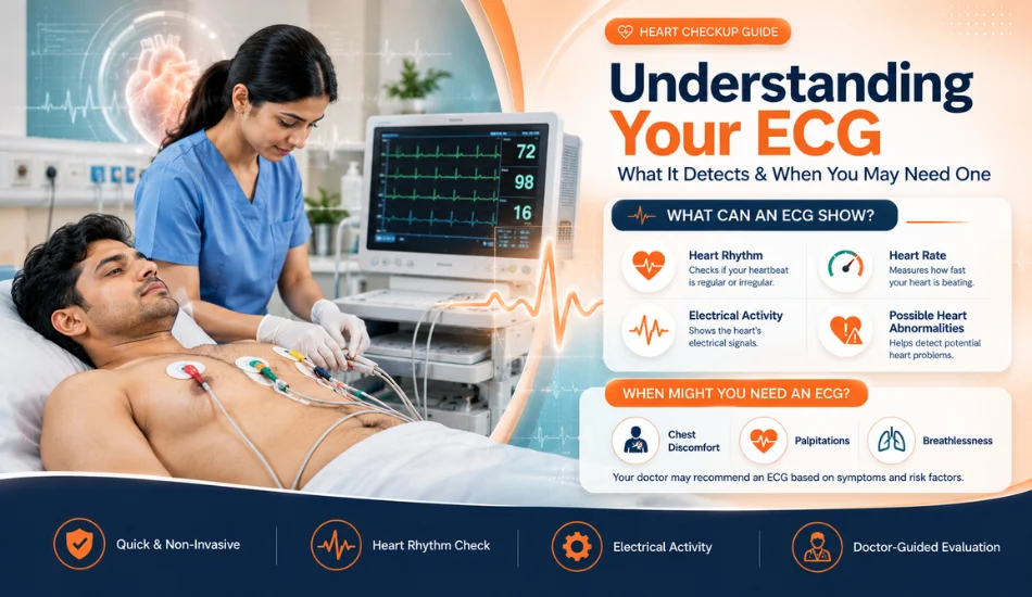
The heart continuously produces electrical signals that coordinate each heartbeat. An electrocardiogram, commonly called an ECG or EKG, is a widely used medical test that records these electrical signals and displays them as a waveform. It is one of the most common investigations used by doctors when evaluating chest discomfort, abnormal heart rhythms, palpitations, dizziness, fainting, or other symptoms that may be related to the heart.
An ECG is quick, non-invasive, and generally painless. It does not involve injections or radiation. Small electrodes are placed on the skin of the chest and limbs to record the heart's electrical activity while the person remains still.
Although an ECG is a simple test, interpreting it correctly requires medical expertise. The result is considered together with symptoms, medical history, physical examination, blood tests, imaging, and other investigations when necessary. A normal ECG does not always rule out every heart problem, while an abnormal ECG does not automatically mean that a person has a serious disease.
Understanding the ECG Test
An electrocardiogram measures the electrical activity generated by the heart during each heartbeat. The electrical signals help the upper and lower chambers of the heart contract in a coordinated manner. An ECG records these signals from different angles and produces characteristic waves and intervals that healthcare professionals can analyze.
Doctors can examine the heart rate, rhythm, timing of electrical conduction, and certain patterns that may suggest heart muscle injury or other cardiac abnormalities. The test can therefore provide valuable information during both routine evaluation and urgent medical assessment.
Table of Contents
1. What Is an ECG Test?
2. How Does an ECG Work?
3. What Does an ECG Detect?
4. Why Might a Doctor Recommend an ECG?
5. ECG for Chest Pain and Heart Symptoms
6. ECG for Abnormal Heart Rhythm
7. What Happens During an ECG?
8. How to Prepare for an ECG
9. Types of ECG Tests
10. Resting ECG vs Exercise ECG
11. What Do ECG Waves Mean?
12. ECG and Heart Attack Evaluation
13. ECG and Arrhythmia Detection
14. Limitations of an ECG
15. FAQs
16. Conclusion
What Does an ECG Detect?
An ECG can provide information about several aspects of cardiac electrical activity. It can identify an abnormal heart rhythm, unusually fast or slow heart rate, certain conduction problems, and patterns that may indicate previous or current damage to the heart muscle.
Depending on the clinical situation, an ECG may help doctors investigate conditions such as atrial fibrillation, some types of heart block, certain abnormal rhythms, and patterns associated with reduced blood flow to the heart. It may also identify electrical changes associated with electrolyte abnormalities or the effects of certain medications.
| What an ECG May Show | Why It Matters |
|---|---|
| Heart Rate | Shows whether the heartbeat is unusually fast, slow, or within an expected range. |
| Heart Rhythm | Can help identify irregular rhythms such as certain forms of arrhythmia. |
| Electrical Conduction | Can reveal delays or abnormalities in how electrical signals travel through the heart. |
| Possible Ischemic Changes | Certain ECG patterns may suggest reduced blood supply to the heart muscle. |
| Previous Heart Damage | Some patterns may be consistent with a previous myocardial infarction or other cardiac injury. |
| Effects of Certain Medicines | Some medications can alter the heart's electrical activity and ECG appearance. |
| Electrolyte-Related Changes | Significant abnormalities in electrolytes can sometimes produce ECG changes. |
Why Might a Doctor Recommend an ECG?
Doctors may recommend an ECG when a person has symptoms that could be related to the cardiovascular system. It can also be performed as part of an evaluation before certain procedures, during monitoring of known heart conditions, or when a doctor wants to investigate a possible abnormal heart rhythm.
Common reasons include chest discomfort, palpitations, unexplained dizziness, fainting, shortness of breath, unusual fatigue, or a sensation of an irregular heartbeat. The decision to perform an ECG depends on the person's symptoms, age, medical history, risk factors, and clinical examination.
| Reason for ECG | What Doctors May Be Evaluating |
|---|---|
| Chest Pain | Possible electrical changes associated with reduced blood supply or heart injury. |
| Palpitations | Irregular, rapid, or abnormal heart rhythms. |
| Dizziness | Possible rhythm or conduction abnormalities that may affect blood flow. |
| Fainting | Possible cardiac rhythm or electrical problems. |
| Shortness of Breath | Possible cardiovascular causes, depending on the overall clinical picture. |
| Known Heart Disease | Monitoring or evaluating changes in cardiac electrical activity. |
| Pre-Procedure Assessment | Assessing cardiac status when clinically appropriate before certain procedures. |
ECG for Chest Pain
Chest pain is one of the important reasons an ECG may be performed. When a person presents with symptoms that could indicate acute coronary syndrome, an ECG can provide rapid information about the electrical activity of the heart.
However, an ECG is only one component of the evaluation. Doctors may also consider symptoms, blood tests such as cardiac biomarkers, physical examination, medical history, and sometimes echocardiography or coronary imaging.
Anyone experiencing sudden or severe chest pressure, especially when accompanied by breathlessness, sweating, nausea, fainting, or pain spreading to the arm, shoulder, back, neck, or jaw, should seek emergency medical attention rather than waiting for a routine appointment.
ECG for Palpitations and Irregular Heartbeat
Palpitations are sensations such as racing, pounding, fluttering, or skipped heartbeats. They can occur for many reasons, including stress, caffeine, certain medications, thyroid problems, anemia, electrolyte abnormalities, or cardiac arrhythmias.
A resting ECG can sometimes identify the rhythm responsible for palpitations. However, if symptoms occur intermittently and the rhythm is normal when the ECG is recorded, a standard test may not capture the abnormality.
In such situations, doctors may recommend longer-term rhythm monitoring, such as a Holter monitor or another ambulatory ECG device, depending on how frequently the symptoms occur.
How Is an ECG Performed?
A standard resting ECG usually takes only a few minutes to record, although preparation and electrode placement may take additional time. The patient normally lies comfortably on an examination bed while small adhesive electrodes are attached to the chest and sometimes the arms and legs.
The electrodes detect the heart's electrical signals. Wires connect the electrodes to the ECG machine, which converts the signals into a series of waveforms. The person generally needs to remain still and avoid talking during the recording because movement can interfere with the tracing.
What Happens Before an ECG?
Before the test, the healthcare professional may ask about symptoms, medications, previous heart conditions, and other relevant medical information. The skin may need to be clean and dry so that the electrodes adhere properly.
In some cases, small areas of hair may need to be trimmed if they interfere with electrode placement. This is usually a simple process and does not require special preparation.
Is an ECG Painful?
A standard ECG is generally painless because it records electrical signals through electrodes placed on the skin. There is no electrical current intentionally delivered into the body during a routine ECG.
Some people may experience minor discomfort when adhesive electrodes are removed, particularly if they have sensitive skin or body hair. Serious complications from a standard resting ECG are uncommon.
How to Prepare for an ECG
Most standard resting ECGs do not require fasting or extensive preparation. Patients should generally follow the instructions provided by their healthcare team.
- Wear clothing that allows easy access to the chest when appropriate.
- Tell the healthcare professional about current medicines and supplements.
- Inform the medical team about known heart conditions.
- Follow any specific instructions provided before the test.
- Remain relaxed and still during the recording.
- Avoid applying excessive lotion or oil to the chest before the test if instructed, as it may interfere with electrode adhesion.
How Long Does an ECG Take?
The actual recording of a resting ECG is usually very quick. Electrode placement and preparation may take longer than the recording itself. In many clinical settings, the complete process can be completed within a short appointment.
The time required for interpretation depends on the clinical setting and whether the ECG is being reviewed immediately for urgent symptoms or as part of a routine assessment.
What Are the Different Types of ECG Tests?
There are several forms of electrocardiographic monitoring. The appropriate type depends on the symptoms and the question the doctor is trying to answer.
| Type | Common Use |
|---|---|
| Resting ECG | Records heart electrical activity while the patient is resting. |
| Exercise ECG | Records the heart's electrical response during controlled physical exertion. |
| Holter Monitoring | Provides continuous ECG recording over an extended period, often to investigate intermittent symptoms. |
| Event Monitoring | May record heart rhythm during episodes of symptoms that occur less frequently. |
| Ambulatory ECG Monitoring | Allows rhythm assessment while the person continues normal daily activities. |
Resting ECG vs Exercise ECG
A resting ECG records electrical activity when the heart is not being deliberately stressed by exercise. An exercise ECG, sometimes called a stress test, records the heart's electrical response while the patient exercises under medical supervision.
Exercise testing may be used in selected patients to investigate symptoms that occur with exertion or to assess cardiovascular response to exercise. It is not appropriate for every person and should be recommended based on clinical assessment.
What Do ECG Waves and Intervals Mean?
An ECG records the electrical activity of the heart as a series of waves and intervals. Doctors examine these patterns to understand how electrical signals move through the heart and whether the heartbeat is regular. Although ECG reports can look complicated, each major wave represents a specific phase of the heartbeat.
P Wave: Electrical Activity of the Atria
The P wave represents the electrical activation of the atria, the two upper chambers of the heart. It occurs before the atria contract and push blood into the ventricles.
Changes in the shape, size, or timing of the P wave may provide clues about an abnormal heart rhythm or changes involving the atria. However, a P-wave abnormality does not by itself establish a specific diagnosis; doctors interpret it along with the complete ECG and the patient's symptoms.
QRS Complex: Electrical Activation of the Ventricles
The QRS complex represents electrical activation of the ventricles, the heart's main pumping chambers. Because the ventricles contain more muscle than the atria, the QRS complex is usually the most prominent part of a normal ECG tracing.
An unusually wide or abnormal QRS complex can sometimes indicate a problem with the electrical conduction system of the heart. It may also be associated with certain heart conditions, previous heart damage, or an abnormal rhythm.
T Wave: Ventricular Recovery
The T wave represents the recovery phase of the ventricles after they have been electrically activated. Doctors look at the T wave and other ECG changes when assessing possible problems such as reduced blood flow to the heart muscle or electrolyte abnormalities.
PR Interval and QT Interval
ECG interpretation also involves measuring specific intervals between waves. The PR interval provides information about the time taken for an electrical signal to travel from the atria toward the ventricles. The QT interval reflects the time required for the ventricles to activate and then recover.
Abnormal intervals can sometimes indicate disturbances in the heart's electrical conduction or an increased risk of certain rhythm problems. Some medications and electrolyte abnormalities can also affect these measurements, which is why the complete clinical situation matters when interpreting an ECG.
| ECG Component | What It Represents | Why Doctors Assess It |
|---|---|---|
| P Wave | Electrical activation of the atria | Helps assess atrial rhythm and electrical activity |
| QRS Complex | Electrical activation of the ventricles | Helps evaluate ventricular conduction and rhythm |
| T Wave | Ventricular recovery | May provide clues about ischemia, electrolyte problems, or other abnormalities |
| PR Interval | Time for electrical conduction from atria toward ventricles | Helps assess conduction through the heart |
| QT Interval | Ventricular activation and recovery | Important when assessing certain electrical rhythm risks |
ECG and Heart Attack Evaluation
An ECG is one of the important tests used when a person has symptoms that could indicate a heart attack. Changes in the ECG can show abnormalities in the heart's electrical pattern that may occur when the heart muscle is not receiving enough blood and oxygen.
Doctors may recommend an ECG urgently when symptoms such as chest pressure, chest pain, unexplained sweating, nausea, shortness of breath, or discomfort spreading to the arm, shoulder, back, neck, or jaw occur.
However, an ECG is not the only test used to evaluate a suspected heart attack. Depending on the situation, doctors may also use cardiac blood tests such as troponin, physical examination, medical history, imaging, and repeated ECGs.
Can an ECG Detect Every Heart Attack?
No. An ECG can provide important information, but a single normal ECG does not completely rule out a heart attack or other serious heart problems. Some abnormalities may develop over time or may not be clearly visible on the first tracing.
When symptoms strongly suggest an acute cardiac problem, doctors may repeat ECG testing and perform additional investigations rather than relying on one normal result.
ECG and Arrhythmia Detection
One of the major uses of an ECG is evaluating abnormal heart rhythms, also called arrhythmias. A standard ECG records the heart rhythm for a short period, allowing doctors to examine heart rate, rhythm regularity, and electrical conduction.
An ECG may help identify or provide clues about rhythm conditions such as atrial fibrillation, certain types of tachycardia, bradycardia, premature beats, and some conduction abnormalities.
| Rhythm Finding | What It May Mean | Possible Symptoms |
|---|---|---|
| Fast heart rate | May occur with certain arrhythmias, fever, stress, dehydration, or other conditions | Palpitations, weakness, dizziness |
| Slow heart rate | May be normal in some people or associated with conduction problems | Fatigue, dizziness, fainting |
| Irregular rhythm | May suggest an abnormal rhythm such as atrial fibrillation | Palpitations, breathlessness, tiredness |
| Premature beats | Extra or early heartbeats that may occur occasionally | Fluttering or skipped-beat sensation |
| Conduction abnormality | Electrical signals may travel through the heart differently than expected | May cause no symptoms or may cause dizziness and other symptoms |
What If Your ECG Is Abnormal?
An “abnormal ECG” does not automatically mean that you have a serious heart disease. ECG machines can identify patterns that require medical review, but the final interpretation depends on symptoms, age, medical history, medications, physical examination, and other test results.
Some ECG changes can be temporary or related to factors such as heart rate, electrolyte levels, medications, previous heart conditions, or technical issues during recording.
If an ECG report mentions an abnormality, the appropriate next step is to discuss the result with a qualified doctor rather than trying to interpret the automated ECG statement independently.
Why Can an ECG Show an Abnormal Result?
Several factors can influence an ECG tracing. These include heart rhythm, electrolyte disturbances, certain medications, previous heart damage, changes in the heart's structure, and problems with electrode placement.
Movement, muscle tension, sweating, poor electrode contact, or incorrect positioning of electrodes can also create artifacts that make the tracing more difficult to interpret.
When a Standard ECG Does Not Capture Symptoms
A standard resting ECG usually records only a short period of heart activity. If palpitations, dizziness, or irregular heartbeats happen only occasionally, the abnormal rhythm may not occur while the ECG is being recorded.
In such cases, a doctor may recommend longer-term monitoring, depending on the frequency and nature of symptoms.
What Is a Holter Monitor?
A Holter monitor is a portable device that continuously records the heart's electrical activity for an extended period, commonly around 24 to 48 hours, although longer monitoring options are also available.
It can be useful when symptoms are intermittent and a short resting ECG does not capture the suspected rhythm abnormality. Patients are generally asked to record symptoms and activities so doctors can compare them with the recorded heart rhythm.
What Is an Exercise ECG or Stress Test?
An exercise ECG records the heart's electrical activity while a person exercises, usually on a treadmill or stationary bicycle under medical supervision. The test allows doctors to observe how the heart responds to increased physical demand.
Depending on the person's symptoms and risk factors, an exercise-based test may help evaluate exercise-related chest discomfort, changes in heart rhythm, blood-pressure response, or other concerns. It is not suitable for everyone, so the decision should be made by a doctor.
Resting ECG vs Exercise ECG vs Holter Monitoring
| Test | Recording Period | Common Purpose |
|---|---|---|
| Resting ECG | Usually a few minutes | Evaluate heart rhythm and electrical activity at rest |
| Exercise ECG | During supervised exercise | Assess heart response to physical exertion |
| Holter Monitoring | Usually 24–48 hours or longer depending on device | Identify intermittent rhythm abnormalities |
ECG Before Surgery
An ECG may sometimes be included in a preoperative assessment, particularly when a patient has certain cardiac risk factors, symptoms, age-related risk, or a medical history that makes cardiac evaluation relevant.
The purpose is not simply to perform the test routinely for everyone. Doctors consider the planned surgery, patient's health status, medications, previous heart conditions, and overall risk before deciding which preoperative investigations are appropriate.
Factors That Can Affect an ECG Recording
For an ECG to provide a useful tracing, electrodes need to be correctly positioned and maintain good contact with the skin. Body movement, talking during the recording, muscle tension, sweating, or loose electrodes can introduce unwanted signals called artifacts.
Medications and changes in electrolyte levels can also influence the ECG. For this reason, doctors interpret the tracing in combination with the patient's medical history and current condition.
When Should You Seek Urgent Medical Attention?
An ECG is often used as part of the assessment of potentially serious heart symptoms. Do not wait for a routine appointment if you develop severe or persistent chest pressure or pain, significant breathing difficulty, fainting, sudden severe weakness, or symptoms suggesting a possible heart attack.
Chest discomfort accompanied by sweating, nausea, shortness of breath, or pain spreading to the arm, shoulder, back, neck, or jaw should be treated as potentially urgent. Seek emergency medical care immediately rather than attempting to arrange a routine ECG yourself.
How Doctors Interpret an ECG Report
ECG interpretation involves much more than reading the words “normal” or “abnormal” printed by the ECG machine. A doctor evaluates the heart rate, rhythm, electrical axis, intervals, waveform patterns, and any significant changes in the context of the patient's symptoms and medical history.
Depending on the findings, further evaluation may include blood tests, echocardiography, stress testing, Holter monitoring, or other cardiac investigations. A single ECG should therefore be considered one part of the overall clinical assessment.
Key Points to Remember About ECG Testing
An ECG is a quick and commonly used investigation that provides valuable information about the heart's electrical activity. It can help doctors evaluate abnormal rhythms, conduction problems, and certain patterns associated with reduced blood supply or previous cardiac injury.
At the same time, an ECG has limitations. A normal result does not guarantee that every heart problem has been ruled out, and an abnormal result does not automatically confirm a serious disease. Proper interpretation requires clinical context and, when necessary, additional testing.
Limitations of an ECG Test
An ECG is a valuable and widely used heart test, but it cannot detect every type of heart problem. A standard ECG records the heart's electrical activity for only a short period. Therefore, some abnormalities may not appear if they occur only occasionally.
An ECG may also provide limited information about the physical structure of the heart. Depending on the symptoms and clinical situation, a doctor may recommend additional tests such as an echocardiogram, stress test, Holter monitoring, blood tests, or other cardiac investigations.
| ECG Limitation | What It Means |
|---|---|
| Short recording time | An intermittent abnormal rhythm may not occur during the test. |
| Cannot assess everything | Some structural or functional heart problems require other investigations. |
| Normal ECG is not a complete guarantee | Some cardiac conditions may still be present despite a normal tracing. |
| Automated interpretation can be imperfect | Computer-generated ECG statements require review by a qualified healthcare professional. |
| Technical artifacts | Movement, poor electrode contact, or incorrect placement can affect the tracing. |
When Might You Need More Than an ECG?
A doctor may recommend additional cardiac tests when symptoms continue despite a normal ECG, when the ECG shows an abnormality, or when your medical history indicates a higher risk of heart disease.
For example, a person experiencing repeated palpitations may benefit from longer heart-rhythm monitoring, while someone with suspected structural heart disease may require an echocardiogram. A person experiencing exertional chest discomfort may be assessed with an appropriate stress-based or imaging test depending on their individual risk.
ECG vs Echocardiogram: What Is the Difference?
An ECG and an echocardiogram are different tests that provide different types of information. An ECG primarily evaluates the heart's electrical activity, while an echocardiogram uses ultrasound to create images of the heart and assess its structure and movement.
| Feature | ECG | Echocardiogram |
|---|---|---|
| Main purpose | Evaluates electrical activity and heart rhythm | Evaluates heart structure, chambers, valves, and pumping function |
| Method | Electrodes attached to the skin | Ultrasound imaging |
| Commonly useful for | Arrhythmias, conduction abnormalities, and certain electrical changes | Valve problems, structural abnormalities, and assessment of heart function |
| Radiation | No | No |
ECG and Blood Tests: Why Both May Be Needed
An ECG and blood tests answer different clinical questions. When a heart attack is suspected, doctors may use an ECG together with cardiac biomarkers such as troponin. The ECG can identify electrical changes, while blood testing can help detect injury to heart muscle.
The timing of symptoms and test results is important. Doctors may repeat tests when clinically necessary because a single test performed at one point in time may not provide the complete picture.
Who May Be Advised to Have an ECG?
An ECG may be recommended for people with symptoms or medical conditions that could involve the heart. It may also be used as part of certain medical evaluations or before selected procedures when a clinician considers it appropriate.
Common situations include:
• Chest pain or pressure
• Palpitations or a sensation of an irregular heartbeat
• Unexplained dizziness or fainting
• Shortness of breath
• Unusually fast or slow pulse
• Suspected arrhythmia
• Previous heart disease
• Assessment of certain cardiovascular risk factors
• Monitoring of some medications that can affect heart rhythm
• Preoperative cardiac assessment when clinically indicated
ECG for People With High Blood Pressure
People with high blood pressure may sometimes undergo an ECG as part of cardiovascular evaluation. Long-standing hypertension can affect the heart, and an ECG may provide clues about certain electrical or structural changes.
However, an ECG does not replace regular blood-pressure monitoring or other assessments recommended by a doctor. Managing blood pressure, cholesterol, diabetes, smoking, weight, physical activity, and other cardiovascular risk factors remains important for overall heart health.
ECG for People With Diabetes
Diabetes is associated with an increased risk of cardiovascular disease. Depending on symptoms, age, medical history, and overall risk, a doctor may consider an ECG or other cardiac evaluation.
People with diabetes should not rely on an ECG alone to assess heart health. Regular medical follow-up and appropriate management of blood sugar, blood pressure, cholesterol, and other risk factors are important.
What Should You Do After an ECG?
In most cases, there is no special recovery period after a routine ECG. Once the electrodes are removed, you can generally return to your normal activities unless your healthcare professional has advised otherwise.
The important step is understanding what the result means in the context of your symptoms. If the ECG was performed because of ongoing symptoms, follow your doctor's recommendations regarding further testing or follow-up.
What If the ECG Report Says “Normal”?
A normal ECG is generally reassuring, but it should be interpreted according to the reason the test was performed. If symptoms such as chest pain, fainting, significant breathlessness, or recurrent palpitations continue, further evaluation may still be necessary.
Do not ignore concerning symptoms simply because an earlier ECG was normal. Heart symptoms can change over time, and some conditions require additional or repeated testing.
What If the ECG Report Says “Abnormal”?
An abnormal ECG should be reviewed by a qualified doctor. Automated ECG systems may flag patterns that require further evaluation, but the machine-generated interpretation is not a substitute for clinical assessment.
An abnormal finding can have several possible explanations, ranging from an incidental variation to a clinically important cardiac condition. Your doctor may compare the ECG with previous recordings and decide whether additional tests are required.
How to Prepare for an ECG Test
A standard resting ECG generally requires little preparation. You can usually eat and drink normally unless your healthcare provider gives different instructions.
Before the test, wear clothing that allows easy access to the chest area. Avoid applying excessive lotions, oils, or creams to the chest on the day of the test because they can interfere with electrode contact.
Tell the healthcare professional about the medicines and supplements you take. Do not stop any prescribed medication unless your doctor specifically instructs you to do so.
Is an ECG Test Painful?
A routine ECG is generally painless because it does not involve needles or electrical shocks being delivered into the body. Small adhesive electrodes are placed on the chest and limbs to record the heart's electrical signals.
Some people may experience mild discomfort when adhesive electrodes are removed, particularly if they have sensitive skin or body hair, but the test itself does not normally cause pain.
How Long Does an ECG Take?
A standard resting ECG is usually completed within a few minutes, although the complete appointment may take longer depending on preparation and clinical assessment.
Longer monitoring tests such as Holter monitoring are different because the device records heart activity over an extended period while you continue your usual activities as instructed.
When Is an ECG an Emergency Test?
An ECG can become part of an urgent assessment when a person develops symptoms that could indicate a serious cardiac problem. Severe or persistent chest pain or pressure, sudden breathlessness, fainting, or other concerning symptoms should not be managed by waiting for a routine test appointment.
If chest discomfort is accompanied by sweating, nausea, difficulty breathing, or discomfort spreading to the arm, shoulder, back, neck, or jaw, seek emergency medical care immediately.
Frequently Asked Questions About ECG Tests
1. What is an ECG test used for?
An ECG records the electrical activity of the heart and helps doctors evaluate heart rate, rhythm, conduction, and certain electrical patterns associated with cardiac conditions. It is commonly used when symptoms such as chest discomfort, palpitations, dizziness, or fainting occur.
2. Can an ECG detect a heart attack?
An ECG can identify electrical changes that may occur during a heart attack and is an important part of emergency cardiac evaluation. However, doctors may also use cardiac blood tests, repeated ECGs, medical history, examination, and other investigations to determine whether a heart attack has occurred.
3. Can an ECG detect an irregular heartbeat?
Yes. An ECG can identify many abnormal heart rhythms when the rhythm disturbance is occurring during the recording. If symptoms are intermittent, a doctor may recommend Holter monitoring or another type of ambulatory heart-rhythm monitoring.
4. Is an ECG safe?
A routine ECG is considered a very low-risk test. It records electrical signals from the heart through electrodes placed on the skin and does not expose the patient to radiation.
5. Does an ECG hurt?
No. A standard ECG is generally painless. Electrodes are attached to the skin, and the machine records the heart's electrical activity. Some people may experience minor discomfort when adhesive electrodes are removed.
6. Can a normal ECG rule out heart disease?
No. A normal ECG can be reassuring, but it cannot rule out every type of heart disease. Some structural problems, intermittent rhythm abnormalities, and other conditions may require additional tests or longer monitoring.
7. What should I do if my ECG is abnormal?
Discuss the report with your doctor. An abnormal automated ECG statement does not necessarily mean that you have a serious heart condition. A qualified healthcare professional should interpret the tracing along with your symptoms, medical history, examination, and other test results.
8. How often should a person get an ECG?
There is no single ECG schedule that is appropriate for everyone. The need for an ECG depends on symptoms, age, cardiovascular risk, medical history, medications, and the reason for evaluation. Your doctor can determine whether and when repeat testing is appropriate.
9. Can an ECG detect blocked arteries?
An ECG may show changes associated with reduced blood flow to the heart, but it does not directly visualize blocked coronary arteries. Depending on the clinical situation, doctors may recommend additional tests to evaluate coronary artery disease.
10. When should I seek immediate medical help instead of waiting for an ECG appointment?
Seek emergency medical attention for severe or persistent chest pain or pressure, significant difficulty breathing, fainting, or chest symptoms associated with sweating, nausea, or pain spreading to the arm, shoulder, back, neck, or jaw. Do not delay emergency care while waiting for a routine ECG appointment.
Conclusion
An ECG is one of the most useful and commonly performed tests for evaluating the heart's electrical activity. It can help doctors assess heart rate and rhythm, identify certain arrhythmias and conduction abnormalities, and provide important clues when evaluating chest pain or suspected cardiac problems.
At the same time, an ECG should not be viewed as a complete test for every type of heart disease. A normal tracing does not rule out all cardiac conditions, while an abnormal result does not automatically mean that a serious disease is present. Doctors interpret ECG findings together with symptoms, medical history, examination, and other investigations when required.
If you experience recurring palpitations, unexplained dizziness, fainting, chest discomfort, unusual breathlessness, or other symptoms that may be related to the heart, consult a qualified healthcare professional. For severe or sudden cardiac symptoms, seek emergency medical care without delay.